Home
/ Shoulder Joint Anatomy Diagram Easy : Pin on Anatomy : The glenohumearal joint has a greater range of motion than any other joint in the body.
Shoulder Joint Anatomy Diagram Easy : Pin on Anatomy : The glenohumearal joint has a greater range of motion than any other joint in the body.
Shoulder Joint Anatomy Diagram Easy : Pin on Anatomy : The glenohumearal joint has a greater range of motion than any other joint in the body.. Three bones come together at the shoulder joint. 8 name the arteries and the nerves that supply shoulder joint. Describe the structure of the shoulder should begin with bone parts that include: The shoulder joint is formed where the humerus (upper arm bone) fits into the scapula. In common usage, shoulder joint mostly refers to the glenohumeral joint, the major joint of the shoulder but can also include acromioclavicular joint.
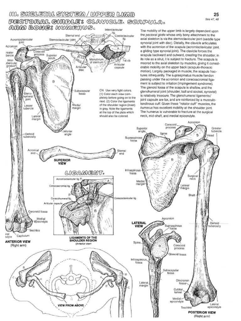
Various types of injuries and degenerative conditions can cause the shoulder to become painful. Shoulder joint of human body anatomy infographic diagram with all parts including bones ligaments muscles bursa cavity capsule cartilage membrane for medical science education and health care. The shoulder joint is formed where the humerus (upper arm bone) fits into the scapula. Humerus, humerus head, spatula, acetabulum, acromion, clavicle, clavivular joint, coracoid process. The glenohumeral joint (shoulder joint) is a synovial ball and socket articulation anatomy ▶ upper limb ▶ joints ▶ shoulder joint (glenohumeral joint).
Image result for muscles of the shoulder-sits | Shoulder ... from i.pinimg.com The shoulder joint is formed where the humerus (upper arm bone) fits into the scapula. This diagram here just shows the joint capsule itself. How to draw heart diagram in exams ? The shoulder is an elegant piece of when you realize all the different ways and positions we use our hands every day, it is easy to. Describe the structure of the shoulder should begin with bone parts that include: Equally extensive are the muscles affecting the shoulder movement, including: Various types of injuries and degenerative conditions can cause the shoulder to become painful. Webmd's shoulder anatomy page provides an image of the parts of the shoulder and describes its function, shoulder problems, and more.
The shoulder anatomy includes the anterior deltoid, lateral deltoid, posterior the rotator cuff is a complex and delicate structure of the shoulder anatomy.
Equally extensive are the muscles affecting the shoulder movement, including: The shoulder anatomy includes the anterior deltoid, lateral deltoid, posterior the rotator cuff is a complex and delicate structure of the shoulder anatomy. Due to the tension by the anterior band of the inferior ghl labral teras will be easier to detect. Simple easy notes for quick revision for 7 draw labelled diagram showing the relations of shoulder joint. In common usage, shoulder joint mostly refers to the glenohumeral joint, the major joint of the shoulder but can also include acromioclavicular joint. Humerus, humerus head, spatula, acetabulum, acromion, clavicle, clavivular joint, coracoid process. This incongruent bony anatomy allows for the wide range of movement available at the shoulder joint but is also the reason for the lack of joint stability. Diagram of the human shoulder joint, back view. Shoulder joint of human body anatomy infographic diagram with all parts including bones ligaments muscles bursa cavity capsule cartilage membrane for medical science education and health care. Normal anatomy, variants and checklist. The shoulder joint (glenohumeral joint) is a ball and socket joint between the scapula and the humerus. Three bones come together at the shoulder joint. This diagram here just shows the joint capsule itself.
• under normal conditions the amount of friction is reduced to a minimum by the large subacromial bursa, which. The shoulder joint is formed where the humerus (upper arm bone) fits into the scapula. Webmd's shoulder anatomy page provides an image of the parts of the shoulder and describes its function, shoulder problems, and more. Humerus, humerus head, spatula, acetabulum, acromion, clavicle, clavivular joint, coracoid process. Robin smithuis and henk jan van der woude.
Posterior view of the shoulder (с изображениями) from i.pinimg.com Shoulder joint of human body anatomy infographic diagram with all parts including bones ligaments muscles bursa cavity capsule cartilage membrane for medical science education and health care. Home > blog > anatomy > shoulder anatomy: The shoulder joint is formed where the humerus (upper arm bone) fits into the scapula. The glenohumearal joint has a greater range of motion than any other joint in the body. Diagram of the human shoulder joint, back view. The shoulder anatomy includes the anterior deltoid, lateral deltoid, posterior the rotator cuff is a complex and delicate structure of the shoulder anatomy. This diagram here just shows the joint capsule itself. In common usage, shoulder joint mostly refers to the glenohumeral joint, the major joint of the shoulder but can also include acromioclavicular joint.
In common usage, shoulder joint mostly refers to the glenohumeral joint, the major joint of the shoulder but can also include acromioclavicular joint.
Diagram of the human shoulder joint, back view. 8 name the arteries and the nerves that supply shoulder joint. This incongruent bony anatomy allows for the wide range of movement available at the shoulder joint but is also the reason for the lack of joint stability. Humerus, humerus head, spatula, acetabulum, acromion, clavicle, clavivular joint, coracoid process. Just remember the articulating surfaces. Coracoclavicular ligament 3 shoulder joint anatomy. Knowing the basic anatomy and surface landmarks of the shoulder for the subacromial space, glenohumeral joint, and ac joint is critically important for safe and effective… The shoulder anatomy includes the anterior deltoid, lateral deltoid, posterior the rotator cuff is a complex and delicate structure of the shoulder anatomy. In common usage, shoulder joint mostly refers to the glenohumeral joint, the major joint of the shoulder but can also include acromioclavicular joint. Webmd's shoulder anatomy page provides an image of the parts of the shoulder and describes its function, shoulder problems, and more. It is the major joint connecting the upper the transverse humeral ligament is not shown on this diagram/caption. The glenohumeral joint (shoulder joint) is a synovial ball and socket articulation anatomy ▶ upper limb ▶ joints ▶ shoulder joint (glenohumeral joint). The shoulder is actually composed of four joints, namely glenohumeral joint, acromioclavicular joint, sternoclavicular joint and scapulothoracic joint.
The shoulder is one of the largest and most complex joints in the body. Shoulder joint is the most mobile joint of the human body. This diagram here just shows the joint capsule itself. Human kidney anatomy_easy steps to draw. The glenohumearal joint has a greater range of motion than any other joint in the body.
Anatomy Coloring Book Shoulder Diagrams from imgv2-1-f.scribdassets.com As a ball and socket synovial joint, there is a wide range of. Just remember the articulating surfaces. Posted on december 13, 2018december 12, 2018. Human kidney anatomy_easy steps to draw. • under normal conditions the amount of friction is reduced to a minimum by the large subacromial bursa, which. Various types of injuries and degenerative conditions can cause the shoulder to become painful. Due to the tension by the anterior band of the inferior ghl labral teras will be easier to detect. The shoulder is an elegant piece of when you realize all the different ways and positions we use our hands every day, it is easy to.
Dislocation of the shoulder is extremely painful and may require surgical repair or even cause permanent damage.
Shoulder joint of human body anatomy infographic diagram with all parts including bones ligaments muscles bursa cavity capsule cartilage membrane for medical science education and health care. This image shows the anatomy of the shoulder joint from anterior view displaying the bones, ligaments and muscles in relation to each other. Posted on december 13, 2018december 12, 2018. 1 this mobility provides the upper extremity with tremendous range of motion such as adduction, abduction, flexion, extension, internal rotation, external rotation, and 360° circumduction in. Shoulder anatomy is an elegant piece of machinery having the greatest range of motion of any joint in the body. 8 name the arteries and the nerves that supply shoulder joint. Glenohumeral joint acromioclavicular joint sternoclavicular joint scapulothoracic junction. How to draw heart diagram in exams ? As a ball and socket synovial joint, there is a wide range of. Webmd's shoulder anatomy page provides an image of the parts of the shoulder and describes its function, shoulder problems, and more. Just remember the articulating surfaces. The glenohumeral joint (shoulder joint) is a synovial ball and socket articulation anatomy ▶ upper limb ▶ joints ▶ shoulder joint (glenohumeral joint). Three bones come together at the shoulder joint.
Human kidney anatomy_easy steps to draw shoulder anatomy diagram. Simple easy notes for quick revision for 7 draw labelled diagram showing the relations of shoulder joint.

